
Fig. 1
- ID
- ZDB-FIG-150430-11
- Publication
- Luz et al., 2014 - Dynamic Association with Donor Cell Filopodia and Lipid-Modification Are Essential Features of Wnt8a during Patterning of the Zebrafish Neuroectoderm
- Other Figures
- All Figure Page
- Back to All Figure Page
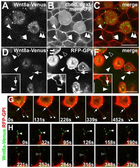
Wnt8a-Venus is membrane associated and released via filopodial processes in the live embryo. Confocal in vivo imaging of Wnt8a sub-cellular localization at the animal pole of 60–90% epiboly embryos. (A–C) Wnt8a-Venus expressing cells colabeled with rhodamine dextran (rhod. dextr., cytoplasmic) transplanted into a host embryo expressing membrane-bound RFP (memRFP). (A) Wnt8a-Venus in producing cells and punctate structures in host tissue (arrowheads). (B) Rhod. dextr.-positive, transplanted cells and memRFP-positive cell membranes of the host embryo. (C) Wnt8a-Venus puncta colocalize with the plasma membrane of surrounding host cells (arrowheads). (D–F) Mosaic coexpression of Wnt8a-Venus and RFP-GPI (labeling plasma membrane). Most Wnt8a-Venus puncta do not colocalize with RFP-GPI (arrowheads), some colocalize with filopodia (arrow). RFP-GPI vesicles do not colocalize with Wnt8a-Venus puncta (black arrowhead). Inset shows magnification of filopodia-associated (arrow) and released (arrowhead) Wnt8a puncta. (A,D) Green channel, (B,E) red channel, (C,F) overlay. Scale bars: 20 µm, 10 µm in inset. (G–H) Single frames of confocal time-lapse series on live embryos with mosaic coexpression of Wnt8a-Venus (green) and RFP-GPI (red). (G) A bifurcated filopodia from Wnt8a producing cell containing two Wnt8a puncta at its tips (arrowheads) and a Wnt8a puncta moving from the cell surface into the filopodia (arrow). (H) Two Wnt8a puncta (white and black arrowheads, respectively) are released from a filopodium of a Wnt8a producing cell. Single confocal planes are shown; time is indicated in seconds (s). |