
Fig. 1
- ID
- ZDB-FIG-150423-23
- Publication
- Williams et al., 2015 - MASH1/Ascl1a Leads to GAP43 Expression and Axon Regeneration in the Adult CNS
- Other Figures
- All Figure Page
- Back to All Figure Page
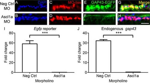
Ascl1a is required for gap43 gene expression in zebrafish. A-H, Cross sections through the retina focused on the retinal ganglion cell layer. A-D, Strong induction of gap43 expression, visualized by the EGFP reporter (C, green; D, yellow), is observed after optic nerve transection in RGCs receiving negative control MOs tagged with lissamine (MO-liss; B, red). In contrast, injury-induced gap43 expression is greatly reduced (G, green; H, yellow) in RGCs receiving Ascl1a MOs tagged with lissamine (F, red). DAPI stained nuclei are blue (scale bar = 5 µm). I, J, Relative fold change over unlesioned control represented graphically. Quantitative real-time PCR demonstrated that there was a reduction in both egfp reporter gene expression (I) and endogenous gap43 expression (J) in gap43:egfp zebrafish that received Ascl1a MOs (n = 4) after optic nerve transection compared to those that received negative control MOs (n = 4; *** = p<0.001, Mann-Whitney T-test). |
| Genes: | |
|---|---|
| Fish: | |
| Condition: | |
| Knockdown Reagent: | |
| Anatomical Terms: | |
| Stage: | Adult |
| Fish: | |
|---|---|
| Condition: | |
| Knockdown Reagent: | |
| Observed In: | |
| Stage: | Adult |