
Fig. 2
- ID
- ZDB-FIG-150423-24
- Publication
- Williams et al., 2015 - MASH1/Ascl1a Leads to GAP43 Expression and Axon Regeneration in the Adult CNS
- Other Figures
- All Figure Page
- Back to All Figure Page
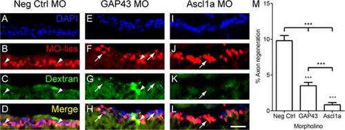
Ascl1a is required for RGC axon regeneration in zebrafish. A-L, Cross sections through the retina focused on the retinal ganglion cell layer. Four days after optic nerve transection, zebrafish RGCs that received MOs (red, arrowheads) were retrogradely traced with dextran (green) 1 mm beyond the site of injury. Animals that received negative control MOs (A-D) were able to regenerate RGC axons as evident by the co-localization of MOs and tracer (yellow, arrow). In contrast few RGCs regenerated axons in animals that received MOs to knockdown GAP43 (E-H) or Ascl1a (I-L). DAPI stained nuclei are blue (scale bar = 10 µm). M, Quantification of the percentage of RGCs that received the MOs and were able to regenerate axons. Compared to controls (n = 6), those that received either GAP43 (n = 6) or Ascl1a (n = 6) MOs exhibited a reduced percentage of RGC axons to regenerate (*** = p<0.001 one-way ANOVA; +++ = p<0.001 Bonferroni posttest). Compared to animals that received GAP43 MOs, those that received Ascl1a MOs exhibited a greater reduction in the percentage of RGC axons to regenerate (*** = p<0.001, t-test). |
| Fish: | |
|---|---|
| Condition: | |
| Knockdown Reagents: | |
| Observed In: | |
| Stage: | Adult |