
Fig. 3
- ID
- ZDB-FIG-150423-25
- Publication
- Williams et al., 2015 - MASH1/Ascl1a Leads to GAP43 Expression and Axon Regeneration in the Adult CNS
- Other Figures
- All Figure Page
- Back to All Figure Page
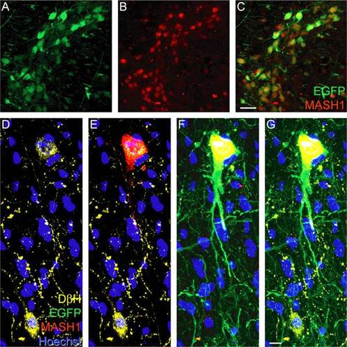
Injection of AAV-EGFP plus AAV-MASH1 induces expression of MASH1 in adult rat brainstem neurons. Immunostaining for EGFP (A, green) and MASH1 (B, red) revealed co-localization in brainstem neurons (C, yellow, scale bar = 20 µm). D, Immunostaining for DβH (yellow) revealed two noradrenergic neurons with Hoechst staining (blue) to visualize nuclei. E, Immunostaining for MASH1 (red) showed that the protein is expressed in the upper DβH-positive neuron. F, MASH1 was only expressed in neurons that also expressed EGFP (green). G, A merged image showed that the lower DβH-positive neuron was not infected with AAV and consequently did not express EGFP or MASH1 (scale bar = 5 µm). |