
Fig. 2
- ID
- ZDB-FIG-150416-8
- Publication
- Cheng et al., 2015 - Nephron proximal tubule patterning and corpuscles of Stannius formation are regulated by the sim1a transcription factor and retinoic acid in zebrafish
- Other Figures
- All Figure Page
- Back to All Figure Page
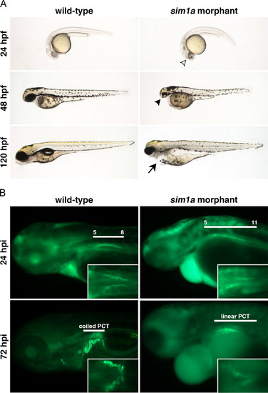
sim1a morphants display drastic developmental defects and abnormal kidney function. (A) A live time course shows the typical morphological phenotypes exhibited by sim1a morphants at different time points – open arrowhead demarcates darkened regions within the head; solid arrowhead indicates small head and eye phenotypes; solid arrow marks the presence of edema. (B) Kidney function in sim1a morphants was analyzed by a 40 kDa dextran-FITC uptake assay where injections were performed at approximately 38 hpf. sim1a morphants displayed partial PCT reabsorption (dorsal inset view) and reduced renal clearance of dextran-FITC over time, with persistent fluorescence throughout the head, trunk and pericardium when compared to wild-types. White lines indicate either the PCT domain in relation to the somites or the PCT morphology at given time points. Embryo anterior is to the left. Abbreviations: hpf – hours post fertilization; hpi – hours post injection; PCT – proximal convoluted tubule. |
| Fish: | |
|---|---|
| Knockdown Reagent: | |
| Observed In: | |
| Stage Range: | Prim-5 to Day 5 |
Reprinted from Developmental Biology, 399(1), Cheng, C.N., Wingert, R.A., Nephron proximal tubule patterning and corpuscles of Stannius formation are regulated by the sim1a transcription factor and retinoic acid in zebrafish, 100-16, Copyright (2015) with permission from Elsevier. Full text @ Dev. Biol.