
Fig. S5
- ID
- ZDB-FIG-150416-17
- Publication
- Cheng et al., 2015 - Nephron proximal tubule patterning and corpuscles of Stannius formation are regulated by the sim1a transcription factor and retinoic acid in zebrafish
- Other Figures
- All Figure Page
- Back to All Figure Page
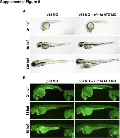
Combined knockdown of p53 and sim1a partially obviates cell death associated with sim1a knockdown. (A) Live time course of embryos injected with a p53 MO or p53+sim1a ATG MO. p53 morphants appear morphologically wild-type. Dual p53+sim1a morphants have wild-type features at 24–48 hpf, but develop pericardial edema and have small head and eye phenotypes at 120 hpf. (B) Cell death was assayed by acridine orange staining. p53 morphants exhibit low levels of cell death as indicated. By comparison, p53+sim1a morphants have slightly higher cell death but this was largely reduced compared to sim1a morphants alone (see Fig. S2A). Embryo anterior is located to the left in all panels. Abbreviations: hpf – hours post fertilization; MO – morpholino. |
Reprinted from Developmental Biology, 399(1), Cheng, C.N., Wingert, R.A., Nephron proximal tubule patterning and corpuscles of Stannius formation are regulated by the sim1a transcription factor and retinoic acid in zebrafish, 100-16, Copyright (2015) with permission from Elsevier. Full text @ Dev. Biol.