
Fig. S4
- ID
- ZDB-FIG-150416-16
- Publication
- Cheng et al., 2015 - Nephron proximal tubule patterning and corpuscles of Stannius formation are regulated by the sim1a transcription factor and retinoic acid in zebrafish
- Other Figures
- All Figure Page
- Back to All Figure Page
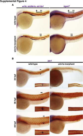
Knockdown of sim1a leads to nephron segmental defects and the loss of the CS. sim1a morphants were screened by WISH at 24 hpf for changes in nephron segmentation. Embryos were assayed with a cocktail of riboprobes to simultaneously detect multiple or single pronephric segments (purple). (A) A mixture of wt1b, slc20a1a and slc12a1 labeled the podocytes, the PCT, and DE, respectively. A single riboprobe was used to directly label the PST (trpm7). (B) WISH analysis of stc1 expression (purple) was performed on sim1a morphants at different time points. Somites are stained in red indicating smyhc1 transcripts. Black bars represent segment domains in correlation to somite numbers. Embryo anterior is to the left in all panels. Abbreviations: CS – corpuscles of Stannius; hpf – hours post fertilization; WISH – whole mount in situ hybridization. |
Reprinted from Developmental Biology, 399(1), Cheng, C.N., Wingert, R.A., Nephron proximal tubule patterning and corpuscles of Stannius formation are regulated by the sim1a transcription factor and retinoic acid in zebrafish, 100-16, Copyright (2015) with permission from Elsevier. Full text @ Dev. Biol.