
Fig. 4
- ID
- ZDB-FIG-150402-21
- Publication
- Wan et al., 2014 - Retinal Injury, Growth Factors, and Cytokines Converge on β-Catenin and pStat3 Signaling to Stimulate Retina Regeneration
- Other Figures
- All Figure Page
- Back to All Figure Page
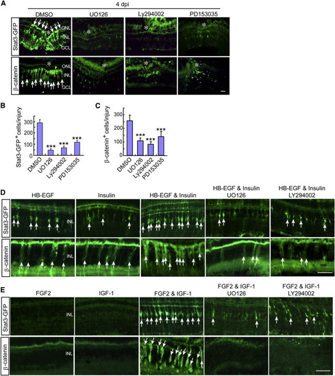
Injury and Growth Factor-Dependent Activation of β-Catenin and Stat3 Signaling in MG-Derived Progenitors Requires Mapk/Erk and PI3K/Akt Signaling (A) Stat3-GFP and β-catenin immunofluorescence shows that inhibition of Mapk (UO126), PI3K (Ly294002), or the Egf receptor (PD153035) suppresses injury-dependent Stat3-GFP and β-catenin accumulation in gfap:stat3-gfp transgenic fish. Asterisks mark the injury site and arrows point to Stat3-GFP+ and β-catenin+ progenitors. Scale bar represents 50 µm. (B and C) Quantification of data shown in (A). Error bars represent SD; p < 0.05; p < 0.001; n = 4. (D and E) Stat3-GFP and β-catenin immunofluorescence shows that intravitreally injected HB-EGF (50 ng) and Insulin (0.5 µg) (D) or FGF2 and IGF-1 (individually at 400 ng or together at 50 ng each) (E) act in a synergistic fashion to stimulate Stat3-GFP and β-catenin accumulation in the uninjured retina of gfap:stat3-gfp transgenic fish and their action is suppressed by inhibition of Mapk (UO126) or PI3K (Ly294002) signaling. Arrows point to Stat3-GFP+ and β-catenin+ progenitors. Scale bar represents 50 µm. |
| Gene: | |
|---|---|
| Fish: | |
| Conditions: | |
| Anatomical Term: | |
| Stage: | Adult |
| Fish: | |
|---|---|
| Conditions: | |
| Observed In: | |
| Stage: | Adult |