|
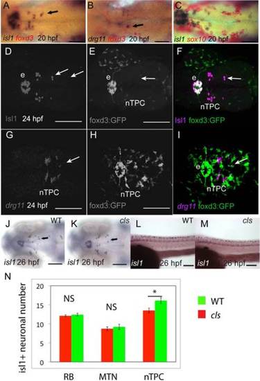
MTN neurons do not arise from neural crest and do not require sox10 function for their development. Expression (blue) of isl1 (A,C) and drg11 (B) in MTN (arrows) and nTPC neurons does not overlap with expression of foxd3 (A,B) and sox10 (C) in mesencephalic neural crest (red) in 24-hpf embryos processed by double in situ hybridisation. Similarly, there is no co-localisation of Isl1 protein (D,F) or drg11 expression (G,I) in MTN neurons with EGFP (E,F,H,I) in neural crest of 24-hpf Tg[foxd3:egfp] embryos, although there is co-localisation in the epiphysis (e). There is no apparent difference in the number of isl1 expressing MTN neurons (J,K) or RB neurons (L,M) in cls mutants (K,M) relative to wildtype (WT) siblings (J,L) at 26 hpf. Quantification of RB, MTN, and nTPC neurons in cls and WT embryos reveals no significant (NS) differences except for a difference in nTPC number (*P<0.05, error bars represent standard error of the mean, N). Scale bars = 100 µm (A–M).
|