
Fig. 6
- ID
- ZDB-FIG-150326-12
- Publication
- Fiengo et al., 2013 - Developmental expression pattern of two zebrafish rxfp3 paralogue genes
- Other Figures
- All Figure Page
- Back to All Figure Page
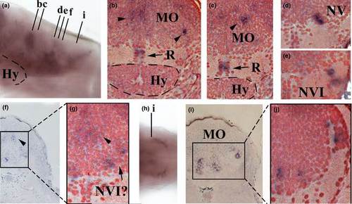
Expression of rxfp3-2b in the posterior region of the 96 h postfertilization (hpf) larval brain. (a) Lateral view of the brain. (b–e) Counter-stained transverse sections as indicated in (a). (f) Transverse section as indicated in (a). (g) Magnification of counter-stained transverse section of the region indicated in (f). (h) Dorsal view of the posterior-most part of the hindbrain. (i) Transverse section as indicated in (h). (j) Magnification of counter-stained transverse section of the region indicated in (i). Black arrowheads indicate cell clusters in the medulla oblongata region. Hy, hypothalamus; MO, medulla oblongata; R, raphe; NV, nuclei of NV cranial nerve; NVI, nuclei of NVI cranial nerve. |
| Gene: | |
|---|---|
| Fish: | |
| Anatomical Terms: | |
| Stage: | Day 4 |