
|
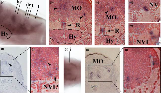
Fig. 6
Expression of rxfp3-2b in the posterior region of the 96 h postfertilization (hpf) larval brain. (a) Lateral view of the brain. (b–e) Counter-stained transverse sections as indicated in (a). (f) Transverse section as indicated in (a). (g) Magnification of counter-stained transverse section of the region indicated in (f). (h) Dorsal view of the posterior-most part of the hindbrain. (i) Transverse section as indicated in (h). (j) Magnification of counter-stained transverse section of the region indicated in (i). Black arrowheads indicate cell clusters in the medulla oblongata region. Hy, hypothalamus; MO, medulla oblongata; R, raphe; NV, nuclei of NV cranial nerve; NVI, nuclei of NVI cranial nerve.