
Fig. 4
- ID
- ZDB-FIG-150313-25
- Publication
- De Smet et al., 2014 - Fibroblast Growth Factor Signaling Affects Vascular Outgrowth and Is Required for the Maintenance of Blood Vessel Integrity
- Other Figures
- All Figure Page
- Back to All Figure Page
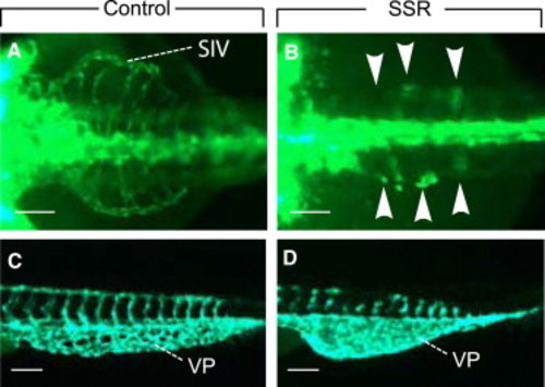
Role of FGFs in Other Angiogenesis Processes in Zebrafish (A and B) Fluorescent images (ventral view) of GFP+ subintestinal vessels (SIVs) on the surface of the yolk sac of 72 hpf Tg(fli1:EGFP)y1 zebrafish embryos, revealing normal SIVs after exposure to DMSO (A) and severe underdevelopment of these SIVs after exposure to 50 μM SSR (tissue concentration 1.9 μg SSR/mg protein) from 20 hpf onward (arrowheads in B). (C and D) Images (lateral view) of the GFP+ vascular plexus (VP) in the posterior trunk of 45 hpf Tg(fli1:EGFP)y1 zebrafish embryos. In DMSO treated control embryos (C), a vascular plexus formed via branching from the caudal vein. After exposure to 200 μM SSR (tissue concentration 3.9 μg SSR/mg protein) from 20 hpfs onward, the vascular plexus failed to remodel and branch and, instead, only a large vascular syncytium was formed (D). In all panels, the head of the embryo faces left; scale bars denote 50 μm in (A) and (B) and 100 μm in (C) and (D). |
| Gene: | |
|---|---|
| Fish: | |
| Condition: | |
| Anatomical Terms: | |
| Stage Range: | High-pec to Protruding-mouth |
| Fish: | |
|---|---|
| Condition: | |
| Observed In: | |
| Stage Range: | High-pec to Protruding-mouth |