
Fig. 4
- ID
- ZDB-FIG-150311-10
- Publication
- Le Pabic et al., 2014 - Fat-Dachsous Signaling Coordinates Cartilage Differentiation and Polarity during Craniofacial Development
- Other Figures
- All Figure Page
- Back to All Figure Page
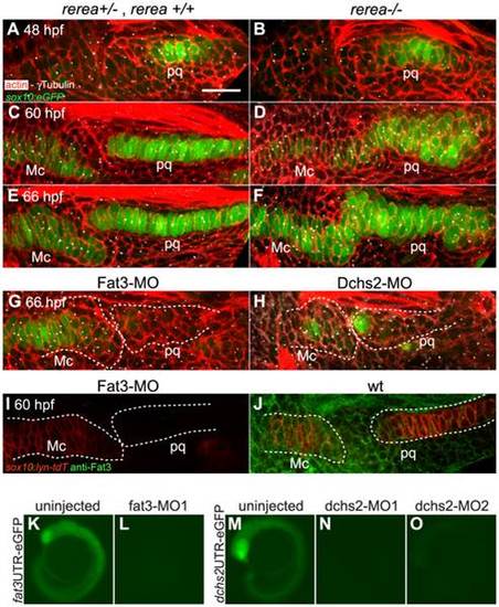
REREa, Fat3 and Dchs2 are required for cartilage stacking and polarity. (A–F): Abnormal stacking and polarity in rerea-/- embryos carrying the sox10:eGFP transgene (green) and stained for cortical actin with phalloidin (red) to reveal cell outlines. Lateral views, anterior to the left. (A, B) Stacking and polarity are comparable in rerea+/+, rerea+/- and rerea-/- pq at 48 hpf. (C, D) Stacking and polarity defects appear in rerea/ pq by 60 hpf, and persist at 66 hpf (E–F). (G, H): Abnormal differentiation, stacking and polarity in Fat3- (G) and Dchs2-deficient embryos (H). (I–J): Fat3-MO injection decreases Fat3 protein levels (loss of green in I) in comparison to WT levels (J). Scale bar = 21µm. (K–O) eGFP reporter RNAs – fat3 52UTR-eGFP (K,L) or dchs2 52UTR-eGFP (M–O) – were injected alone or together with either fat3-MO1 (L), dchs2-MO1 (N) or dchs2-MO2 (O), and eGFP fluorescence measured at 18–20 hpf. Views are lateral, with anterior to the left. Mc: Meckel′s; pq: palatoquadrate. |
| Gene: | |
|---|---|
| Fish: | |
| Knockdown Reagents: | |
| Anatomical Terms: | |
| Stage Range: | Long-pec to Pec-fin |
| Fish: | |
|---|---|
| Knockdown Reagents: | |
| Observed In: | |
| Stage Range: | Long-pec to Pec-fin |