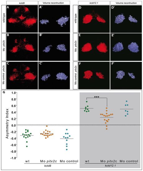
Partial left isomerisation in pitx2c morphants. (A-C) Single confocal sections of the epithalamus showing in situ hybridisation against kctd8 in wild-type, pitx2c morphant and control morphant embryos at 72 hpf. No differences are detected between the different contexts. (A2-C2) Three-dimensional renderings of volumes of kctd8 expression in the habenulae of the respective confocal acquisition sets. (D-F) Single confocal sections of the epithalamus showing in situ hybridisation against kctd12.1 in wild-type, pitx2c morphant and control morphant embryos at 72 hpf. pitx2c morphants display broader expression of kctd12.1 in the right habenula than either of the control contexts. (D2-F2) Three-dimensional renderings of volumes of kctd12.1 expression in the habenulae of the respective confocal acquisition sets. (G) Asymmetry index (AI) with regard to the volumes of kctd8 or kctd12.1 expression for individual wild-type (wt), control or pitx2c morphant embryos. Horizontal black line represents the median AI for each context and the dotted line indicates symmetry. Only the AI for the expression of kctd12.1 in pitx2c morphants is significantly more symmetric than control conditions; ***P<0.001 using a t-test. Embryos are viewed dorsally with the anterior to the top.
|
