
Fig. S2
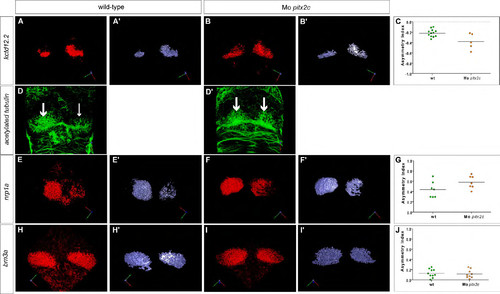
(A, B) Single confocal sections of the epithalamus showing in situ hybridisation against kctd12.2 in wild-type and pitx2c morphant embryos at 72 hpf. (A′, B′) 3D renderings of volumes of kctd12.2 expression in the habenulae of the respective confocal acquisition sets. (C) Asymmetry index (AI) with regard to the volume of kctd12.2 expression for individual wild-type or pitx2c morphant embryos. Horizontal black line represents the median AI for each context: note that the AI is negative because the asymmetry is biased to the right. No significant difference between wild-type or pitx2c morphant embryos is detected. (D, D′) Single confocal sections through the epithalamus showing the habenular neuropil detected by immunostaining against acetylated tubulin at 72 hpf. While the neuropil of wild-type embryos is asymmetric with a left bias (large versus small arrows), symmetric anti-acetylated tubulin staining is detected in pitx2c morphant embryos (two large arrows). (E, F) Single confocal sections of the epithalamus showing in situ hybridisation against nrp1a in wild-type and pitx2c morphant embryos at 72 hpf. (E′, F′) 3D renderings of volumes of nrp1a expression in the habenulae of the respective confocal acquisition sets. (G) Asymmetry index with regard to the volume of nrp1a expression for individual wild-type or pitx2c morphant embryos. Horizontal black line represents the median AI for each context. No significant difference between wild-type or pitx2c morphant embryos is detected. (H, I) Single confocal sections of the epithalamus showing in situ hybridisation against brn3a in wild-type and pitx2c morphant embryos at 72 hpf. (H′, I′) 3D renderings of volumes of brn3a expression in the habenulae of the respective confocal acquisition sets. (J) Asymmetry index with regard to the volume of brn3a expression for individual wild-type or pitx2c morphant embryos. Horizontal black line represents the median AI, which are in both contexts close to zero reflecting that they are virtually symmetric. No significant difference between wild-type or pitx2c morphant embryos is detected. Embryos are view dorsally with the anterior to the top. |