
Fig. S2
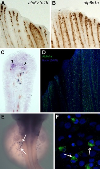
V-ATPase subunits localization in chloride cells, intact and regenerating fins. Whole mount in situ hybridization for atp6v1e1b (Figure A) and atp6v1a (Figure B) in intact fins. Cross section of the whole mount in situ hybridization for atp6v1a at 24 hours post amputation (hpa) where expression can be observed in the blastema, distal to the bone (Figure C, arrowheads). Atp6v1a in the intact caudal fin is present mainly in the epidermis, in a scattered pattern (Figure D). Whole mount in situ hybridization (Figure E) and immunostaining (Figure F) for atp6v1a subunit in the chloride cells of the zebrafish embryo. |