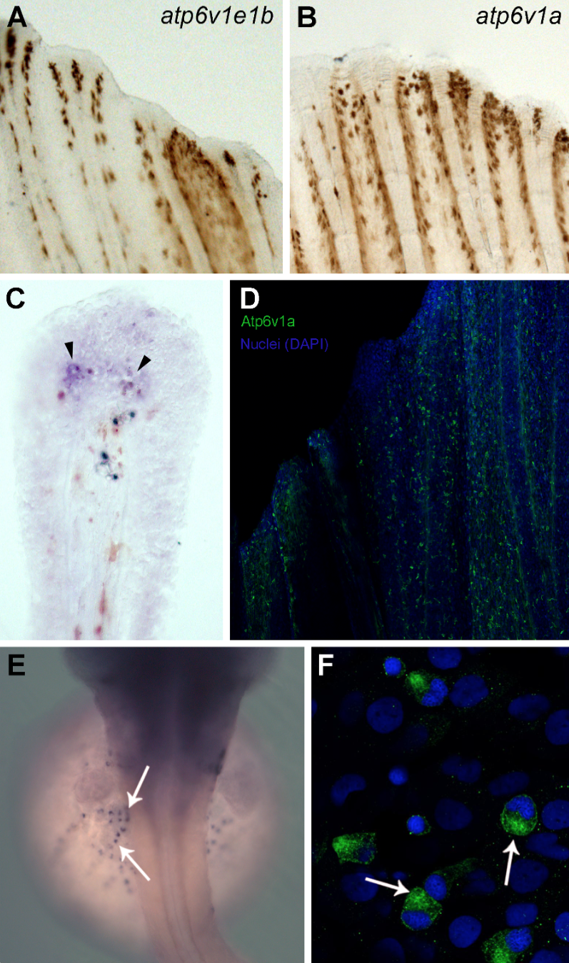
Fig. S2 V-ATPase subunits localization in chloride cells, intact and regenerating fins. Whole mount in situ hybridization for atp6v1e1b (Figure A) and atp6v1a (Figure B) in intact fins. Cross section of the whole mount in situ hybridization for atp6v1a at 24 hours post amputation (hpa) where expression can be observed in the blastema, distal to the bone (Figure C, arrowheads). Atp6v1a in the intact caudal fin is present mainly in the epidermis, in a scattered pattern (Figure D). Whole mount in situ hybridization (Figure E) and immunostaining (Figure F) for atp6v1a subunit in the chloride cells of the zebrafish embryo.
Image
Figure Caption
Acknowledgments
This image is the copyrighted work of the attributed author or publisher, and
ZFIN has permission only to display this image to its users.
Additional permissions should be obtained from the applicable author or publisher of the image.
Full text @ PLoS One

