
Fig. 2
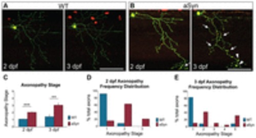
Early axonopathy in aSyn-expressing peripheral sensory neurons. Axon pathology was scored at 2 and 3 days post-fertilization (dpf) in WT and aSyn-expressing axons, using the 5-point staging system described in supplementary material Fig. S3. (A) At 2 dpf (48-58 hpf) and 3 dpf (72-82 hpf), WT axons were smooth and continuous (score of 1). (B) At 2 dpf, axonal beading (arrows) was observed in many aSyn-expressing cells. By 3 dpf, axonal dystrophy in aSyn-expressing cells was more severe, with more diffuse beading (arrows) and larger varicosities (arrowheads). (C) Quantification of average axonopathy stage in wild-type and aSyn-expressing embryos. aSyn-expressing axons were more dystrophic at both 2 dpf (WT degeneration stage: 1.03±0.08, n=12 axons in 5 animals; aSyn: 2.05±0.14; n=19 axons in 8 animals; ***P<0.0001) and 3 dpf (WT: 1.42±0.34; aSyn: 3.05±0.35; ne12 axons in e5 animals as above; **P=0.0033). (D,E) Histograms representing frequency distribution of axonopathy stage. At 2 dpf (D), 11/12 wild-type axons (91.7%) were smooth and continuous (stage 1); one exhibited mild beading (stage 2). By contrast, only 3/19 (15.8%) aSyn-expressing axons were at stage 1; 12/19 (63.2%) exhibited mild beading (stage 2), and 4/19 (21.1%) exhibited more severe axonopathy (stage 3). (E) Frequency histogram of axonopathy distribution in the same cells at 3 dpf. One wild-type axon (8.3%) exhibited mild beading (stage 2), and one wild-type cell had undergone developmental cell death (stage 5). All remaining wild-type axons (10/12, 83.3%) were smooth and continuous (stage 1). By contrast, only 2/19 (10.5%) aSyn-expressing axons remained at stage 1 by 3 dpf. 6/19 (31.6%) had fully degenerated (stage 5), and the remaining 11/19 (57.9%) were in intermediate stages of degeneration (8/19 in stage 2; 2/19 in stage 3; 1/19 in stage 4). Scale bars: 100 μm. |