
Fig. 4
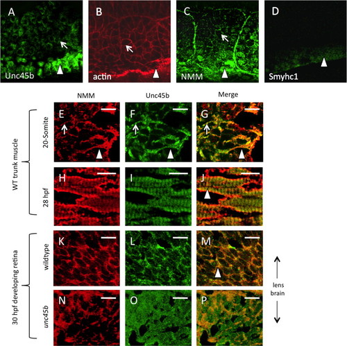
Unc45b protein is co-localized with non-muscle myosin protein in early paraxial mesoderm. Immunofluorescent staining of 5-somite (A–D) zebrafish embryos demonstrates protein accumulation of Unc45b (A) and non-muscle myosin (C) in both adaxial cells (arrowheads) and lateral paraxial mesoderm, demonstrating enrichment at the myoblast cortex (arrows) and somite boundaries, where cytoskeletal actin is also enriched (B). By contrast, muscle myosin protein accumulation (D) was limited to adaxial cells that form the superficial layer of slow muscle fibers. In cryosections of developing zebrafish muscle tissue (E–J), partial co-localization of Unc45b (green) and NMM (red) protein can be seen as yellow fluorescence within myoblast cortices (panels G and J). At the 20-somite stage (E–G), localization to myoblast cortices (arrows) and nascent myofibrils (arrowheads) are both evident. This partial co-localization persists at 28 hpf (H–J). In developing retinal tissue near the lens, Unc45b (K) and NMM (L) can also be seen to co-localize at 48 hpf (yellow fluorescence in panel M); however, this co-localization was lost in unc45b mutant embryos lacking the C-terminal myosin-binding UCS domain (N–P). Merged NMM and Unc45b staining is shown in panels G, J, M and P. Scale bars=10 µm. |
| Gene: | |
|---|---|
| Antibodies: | |
| Fish: | |
| Anatomical Terms: | |
| Stage Range: | 5-9 somites to Long-pec |
Reprinted from Developmental Biology, 390, Myhre, J.L., Hills, J.A., Jean, F., Pilgrim, D.B., Unc45b is essential for early myofibrillogenesis and costamere formation in zebrafish, 26-40, Copyright (2014) with permission from Elsevier. Full text @ Dev. Biol.