
Fig. 1
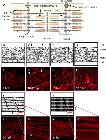
Diagrammatic representation of zebrafish somite maturation during myogenesis. (A) Schematic representation of the sarcomere in skeletal muscle, labeled to show the major protein components (modified from (Myhre and Pilgrim, 2012)). Panels B–E, J and K represent a dorsal view of paraxial mesoderm extending laterally from the edge of the developing neural tube (only the mesoderm is shown). Sample embryos at each stage are stained with phalloidin to show actin dynamics (F–I, L–O). Presomitic mesoderm in early embryos is made up of disorganized proliferative cells with thickened cortical actin structures (B, F). A columnar epithelium can be found immediately adjacent to the developing neural tube; these are the early adaxial cells. (C) Adaxial cells undergo apical constriction as the cells of the segmental plate align to create continuous actin barriers between the somites (arrowheads in C, G), and somites first become visible within the embryo. Fusion of adaxial cells results in the formation of the first contractile structures (D, H) containing slow-fiber myosin isoforms. Clockwise rotation of the somites begins at approximately the 15-somite stage, creating a curvature of the myosepta (E, I) that gives rise to the characteristic chevron shape. Concurrently, adaxial slow muscle fibers migrate laterally towards the somite periphery (arrow in I). At this stage, somite cells of the lateral paraxial mesoderm begin to elongate and fuse. Once somite cells are fully fused (J, L), striations appear in the cortical actin as the first myofibrils are constructed (arrows). As myocytes mature, they fill completely with striated myofibrils (K, N). Panels M and O show the formation of striated myofibrils at higher magnification. All artwork by Alina Pete. |
Reprinted from Developmental Biology, 390, Myhre, J.L., Hills, J.A., Jean, F., Pilgrim, D.B., Unc45b is essential for early myofibrillogenesis and costamere formation in zebrafish, 26-40, Copyright (2014) with permission from Elsevier. Full text @ Dev. Biol.