
Fig. 3
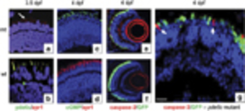
Expression of cell death markers in the retinal sections. (a, c, e, g) Mutant pde6c (mt) retinal images. (b, d, f) Wild-type (wt) retinal images. (a and b) Double immunostaining with zpr1 cone-specific marker in red and pde6c in green. (c and d) Double immunostaining for cGMP labelling in green and zpr1 in red. (e and f) Activated caspase-3 localization in red on GFP-expressing cone transgenic background. (g) Higher magnification of retina in image (e) showing no overlap of caspase-3 red staining in GFP-expressing cones (green). All sections counterstained with DAPI. Scale bar is 5μm, except in e and f, where it is 20μm |
| Gene: | |
|---|---|
| Antibody: | |
| Fish: | |
| Anatomical Terms: | |
| Stage Range: | Protruding-mouth to Day 4 |