|
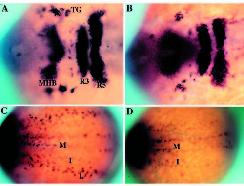
Phenotypic basis for isolation of nrd during the genetic screen. Dorsal views of 2-3 somite stage haploid embryos (anterior to the right) upon analysis by in situ hybridization utilizing a combination of RNA probes: her5 (to visualize the midbrain hindbrain boundary), Krox20 (to visualize rhombomeres 3 and 5) and HuC (to visualize trigeminal ganglia (TG) and medial (M), intermediate (I) and lateral (L) domains of primary neurons). Wild type (A,C) and nrd (B,D) embryos with view of anterior (A,B) and trunk neural plate (C,D) region. nrd has normal anterior-posterior patterning but lacks the most lateral stripe of primary neurons.
|