
Fig. 1
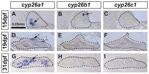
In zebrafish, cyp26a1 is the main Cyp26 paralog expressed in gonads during the critical period for sex-determination. Undifferentiated gonads at 15 days post-fertilization (dpf) expressed cyp26a1 (A) and cyp26b1 (B), but did not express cyp26c1 at detectable levels (C) (A-C: n=4). In bipotential gonads at 19 dpf, cyp26a1 expression became restricted mainly to the dorsal margin of the gonad (D) but expression of cyp26b1 and cyp26c1 was not detected (E, F) (D–F: n=9). In differentiating testes at 31 dpf, cyp26a1 expression up-regulated (G) and in contrast to mouse testes, which up-regulate cyp26b1, neither cyp26b1 nor cyp26c1 expression was detected in maturing zebrafish gonads (H, I) (G-I: n=10). Differentiating testes were assigned by morphological features and assessed by the expression of the male specific Amh marker (see Figure 4). We conclude that in zebrafish, Cyp26a1 is expressed at the time and place necessary to provide an RA-degrading function equivalent to Cyp26b1 in tetrapods. These results suggest independent subfunction partitioning of ancestral cyp26 regulatory elements in lineages leading to zebrafish and mouse. Arrows point to examples of expressing cells. Dashed lines outline gonads. Scale bar: 0.05mm. |
| Genes: | |
|---|---|
| Fish: | |
| Anatomical Terms: | |
| Stage Range: | Days 14-20 to Days 30-44 |