
Fig. 2
- ID
- ZDB-FIG-130726-28
- Publication
- Head et al., 2013 - Activation of canonical Wnt/beta-catenin signaling stimulates proliferation in neuromasts in the zebrafish posterior lateral line
- Other Figures
- All Figure Page
- Back to All Figure Page
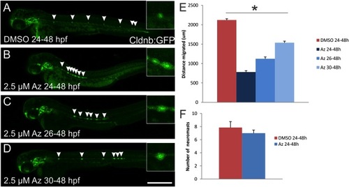
Pharmacological inhibition of GSK3β with Az limits migration of the lateral line primordium. Whole-mount confocal examination of Tg(cldnb:gfp) zebrafish at 48hpf shows the extent of lateral line primordium migration and the location of deposited neuromasts (arrowheads). A: Embryos treated with DMSO as a vehicle control from 24–48 hpf deposit 7–9 neuromasts extending from the ear to the end of the tail. B: Embryos treated with 2.5 μM Az from 24–48 hpf show greatly reduced migration of the primordium, though 7–9 neuromasts were still deposited. C,D: Delaying the onset of Az treatment to 26 hpf (C) or 30 hpf (D) allowed the primordium to migrate further down the tail, but had little effect on the number of deposited neuromasts. In addition to the distance migrated, the size of the neuromasts in Az-treated fish appears larger than controls (insets show magnified view of the central-most neuromast for each fish). E: Quantification of the distance along the lateral midline beginning at the otic vesicle that the primordium had migrated by 48 hpf (n = 6 embryos per condition) in fish treated at varying times with Az or DMSO as a control. All pairwise comparisons between conditions are statistically significant (P < 0.05; ANOVA with post-hoc Tukey). F: Quantification of the number of deposited neuromasts at 48 hpf in fish treated with DMSO or Az at 24 hpf. Though Az significantly reduced the migration of the primordium, there was no significant effect on the number of deposited neuromasts (P > 0.05; t-test). Scale bar = 500 μm. |