
Fig. 7
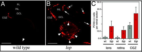
Cell proliferation in the lens and retina. Proliferating cell nuclear antigen (PCNA) immunolabeling detected proliferating cells in the retina and lens. A: A few PCNA-positive cells at the retinal margin (circumferential germinal zone [CGZ]) and a single cell in the lens epithelial cell layer of a wild-type eye (arrowhead). B: Large numbers of PCNA-labeled cells in the lop mutant lens (arrowheads) and retina. In the mutant retina, the PCNA-positive cells are located within the CGZ and the distal (arrows) and proximal (double arrows) aspects of the inner nuclear layer. C: The PCNA-positive cells were quantified in the wild-type and lop mutant lens, retina, and CGZ. Error bars show SD (n values, see Experimental Procedures section and Table 1). PL, photoreceptor layer; INL, inner nuclear layer; GCL, ganglion cell layer; CGZ, circumferential germinal zone; wt, wild-type. Scale bars = 50 µm in A,B. |