
Fig. 2
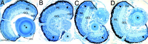
Histological analysis. A-D: Three-micrometer plastic sections were stained with methylene blue and azure II. A: In the 7 days postfertilization wild-type eye, the three retinal layers, which include the ganglion cell (GCL), inner nuclear (INL), and photoreceptor layer (PL) can be identified. The circumferential germinal zone (CGZ) is located at the peripheral margin of the retina. In addition, the wild-type lens epithelial cells (LEC), cortex (C), and nucleus (N) are evident. B-D: Three different lop mutant eyes are shown. The lens structure is severely altered in all three fish, with the putative nucleus labeled by an asterisk. In addition, the mutant retinas lack a distinct photoreceptor layer, although groups of photoreceptors may exist at the periphery (arrowheads). Scale bar = 50 µm. |
| Fish: | |
|---|---|
| Observed In: | |
| Stage: | Days 7-13 |