
Fig. 5
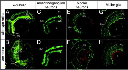
Retinal structure and cell type identification in wild-type and lop mutants. Wild-type (A,C,E,G) and lop mutant retinas (B,D,F,H) were stained with different antibodies to examine the retinal organization. A,B: The α-tubulin antiserum, which stains a component of the retinal cell cytoskeleton, shows a nearly identical pattern in the wild-type and lop mutant (A and B, respectively). B: The major difference is the presence of the wild-type photoreceptor layer that is significantly reduced in the lop mutant (arrow). C,D: The amacrine and ganglion cells, which were identified by immunolocalization of the RNA-binding protein HuC/D, are largely identical in wild-type (C) and mutant (D) retinas. E,F: The subset of retinal bipolar cells that are stained with the anti-PKC are equivalent in wild-type and lop (E and F, respectively). G,H: Müller cells, which were identified by glutamine synthetase detection, were also equivalent in the wild-type (G) and mutant (H) retinas. PL, photoreceptor layer; INL, inner nuclear layer; GCL, ganglion cell layer; IPL, inner plexiform layer; OPL, outer plexiform layer; OpN, optic nerve; L, lens. Scale bar = 50 µm. |