
Fig. 4
- ID
- ZDB-FIG-130502-18
- Publication
- Song et al., 2013 - Pou5f1-dependent EGF expression controls e-cadherin endocytosis, cell adhesion, and zebrafish epiboly movements
- Other Figures
- All Figure Page
- Back to All Figure Page
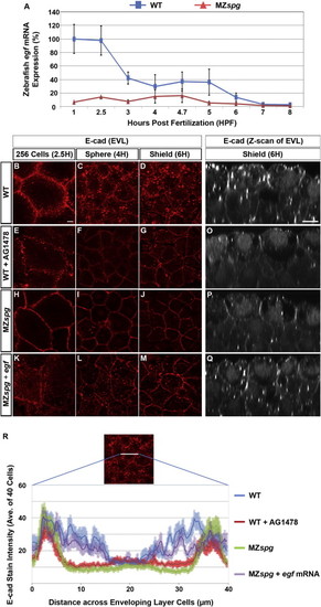
Pou5f1 May Control E-cad Intracellular Localization via Regulation of EGF Expression(A) Time series microarray data of egf mRNA expression in WT and MZspg embryos from four-cell stage to 75% epiboly (1–8 hpf; Onichtchouk et al., 2010). From 1 hpf to MBT, p < 0.01 (n = 3, error bars show SD).(B–Q) E-cad subcellular localization at 256 cells, sphere, and shield stage: (B–M) confocal planes of whole mount anti-E-cad immunofluorescence. Animal views. (N–Q) Confocal z-sections (50 μm) of anti-E-cad ECD immunofluorescence whole mount embryos. Lateral views, animal pole to the top. Scale bars, 10 μm.(R) Quantification of E-cad immunofluorescence signal in a line (highlighted in image) across individual EVL cells. The plot profile represents intensity of E-cad stain in EVL cells of WT, AG1478-treated WT, MZspg, or egf mRNA-injected MZspg embryos (average of 40 cells for each condition). Error bar profiles show SEM. |
| Antibody: | |
|---|---|
| Fish: | |
| Condition: | |
| Anatomical Terms: | |
| Stage Range: | 256-cell to Shield |
Reprinted from Developmental Cell, 24(5), Song, S., Eckerle, S., Onichtchouk, D., Marrs, J.A., Nitschke, R., and Driever, W., Pou5f1-dependent EGF expression controls e-cadherin endocytosis, cell adhesion, and zebrafish epiboly movements, 486-501, Copyright (2013) with permission from Elsevier. Full text @ Dev. Cell