|
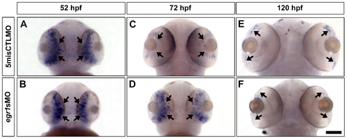
The expression of ptf1a, a TF that specifies ACs and HCs, was abnormal in the Egr1-morphant retinas. Whole-mount in situ hybridization of ptf1a was performed with the controls (5misCTLMO) and Egr1 morphants (egr1sMO) collected at 52, 72 and 120 hpf. At 52 hpf, ptf1a was primarily expressed in the differentiating retinal neuroepithelium (A & B, arrows) in both types of samples. By 72 hpf, the expression of ptf1a was restricted to the proliferative MZ in the controls (C, arrows), while its expression was maintained in the developing central retina in the Egr1 morphants (D, arrows). This ectopic expression was transient, as ptf1a was finally expressed in MZ in the Egr1 morphants (F, arrows) in a very comparable manner as the controls (E, arrows). The ventral view of the embryos is shown in all pictures. Scale bar = 100 µm.
|