
Fig. 4
- ID
- ZDB-FIG-130320-30
- Publication
- Choi et al., 2013 - In vivo monitoring of cardiomyocyte proliferation to identify chemical modifiers of heart regeneration
- Other Figures
- All Figure Page
- Back to All Figure Page
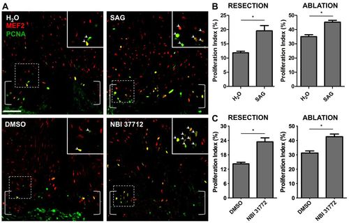
Hedgehog or Igf pathway activation increases cardiomyocyte proliferation during regeneration in zebrafish. (A) Treatment with SAG (2.5 μM) or NBI-31772 (10 μM) from 6 to 7 dpa increased cardiomyocyte proliferation. Mef2, red; PCNA, green. Brackets indicate injury site. Insets: High magnification of the boxed areas. Arrowheads indicate proliferating cardiomyocytes. Scale bar: 50 μm. (B) Quantification of cardiomyocyte proliferation following treatment with SAG (2.5 μM) after resection (left) or ablation (right) injury models. Fish were treated from 6 to 7 dpa. n=9-15, mean±s.e.m. *P<0.01, Student′s t-test. (C) Quantification of cardiomyocyte proliferation following treatment with NBI-31772 after resection (10 μM) or ablation (5 μM) injury models. n=11-17, mean±s.e.m. *P<0.05, Student′s t-test. |