FIGURE

Fig. 2

ID
ZDB-FIG-130220-12
Publication
Luo et al., 2012 - Midkine-A functions upstream of Id2a to regulate cell cycle kinetics in the developing vertebrate retina
Other Figures
All Figure Page
Back to All Figure Page
Fig. 2

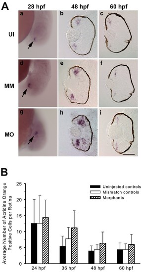
Loss of Mdka function does not affect neurogenic competence or increase cell death. (A) In situ hybridizations showing atoh7 expression in the retinas of uninjected (a-c), control (d-f) and Mdka loss-of-function embryos (g-i). Arrows in panels a, d and g identify the initial cluster of atoh7-expressing cells in ventral retina. MM, embryos injected with 5-nucleotide mismatch morpholinos; MO, embryos injected with mdka-targeted morpholinos; UI, uninjected. Scale bar equals 50μm. (B) Average number of acridine orange-stained cells in the retinas of control and experimental embryos showing that loss of Mdka does not increase cell death. P >0.05 for all pairwise comparisons. n=5 to 7 embryos/time point. Scale bar equals 50 μm.

Expression Data
Gene:
Fish:
Knockdown Reagent:
Anatomical Terms:
Stage Range: Prim-5 to Pec-fin

Expression Detail
Antibody Labeling
Phenotype Data
Fish:
Knockdown Reagent:
Observed In:
Stage Range: Prim-5 to Long-pec

Phenotype Detail
Acknowledgments
This image is the copyrighted work of the attributed author or publisher, and ZFIN has permission only to display this image to its users. Additional permissions should be obtained from the applicable author or publisher of the image. Full text @ Neural Dev.