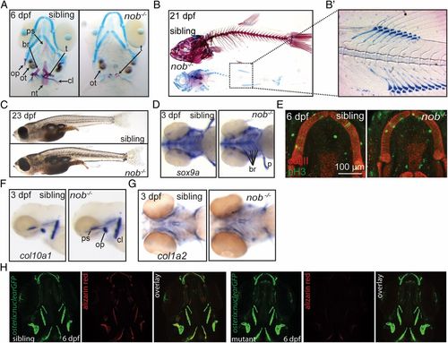
Nob mutants lack a mineralized skeleton. (A) Alizarin red/alcian blue staining of sibling and mutant nob embryos. Cartilage elements appear normal in mutants. All bone is absent, but teeth and otoliths are present. (B) Skeletal staining of 21-dpf sibling and nob mutant individuals. (B2) Enhanced contrast image highlighting the correctly patterned but unmineralized vertebra anlagen. (C) Images of sibling and mutant nob fish at 23 dpf, demonstrating that nob mutants are indistinguishable from siblings at the gross morphological level. (D) Whole-mount in situ hybridization detecting the chondrogenic marker sox9a. (E) Confocal projection of Meckel’s cartilage of a sibling versus mutant embryo showing no difference for anti-type II collagen (red) or the proliferation marker anti–phospho-Histone H3 (green). (F and G) Whole-mount in situ hybridization detecting the osteoblast markers col10a1 (F) and col1a2 (G) in 3-dpf nob mutant and sibling embryos. (H) Osterix:GFP expression, marking early osteoblasts in 6-dpf nob mutant and sibling embryos. br, fifth branchial arch; cl, cleithrum; nt, notochord tip; op, operculum; ot, otolith; ps, parasphenoid; t, teeth.
|
