Expression of Nonvisual Opsins in the otpa Domain and Expression of opn4a in Wild-Type and otp Mutant Embryos, Related to Figure 3
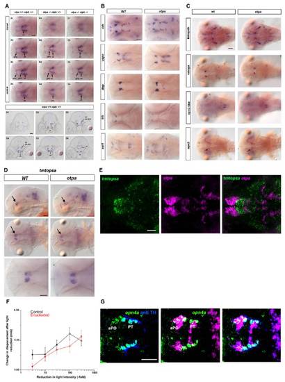
(A) Expression on opn4a in wild-type and otpa mutant embryos. opn4a expression was analyzed in 3dpf larvae by whole-mount in situ hybridization. Top panels: dorsal views of individual focal planes of the larval head in dorsal to ventral order. The otp mutant genotypes are indicated at top. otpa and otpa otpb double mutant larvae reveal loss of opn4a expression in the aPO (anterior preoptic region) and PT (posterior tuberculum). pPO-posterior preoptic region, vHventral hypothalamus. Dorsal views. Scale bar is 50 μm. Bottom panels: frontal sections of a wild-type larval head in rostral to caudal order reveal the anatomical position of opn4a expressing neurons (see also [2]). ac-anterior commissure; aPO-anterior preoptic region; cHcaudal hypothalamus; DA DC2 / DA DC4 - Otp dependent posterior tubercular dopaminergic groups DC2 (rostral) and DC4 (caudal); PT-posterior tuberculum; vH-ventral hypothalamus. Frontal views, dorsal at top. Scale bar is 50 μm.
(B) Presence of neurohormone producing cells in the preoptic region in wild-type and otpa mutants. Expression of crh, vsnp, itnp, trh, and sst1 at 3dpf in wild-type embryos and otpam866 mutants. Whole-mount in situ hybridization (3dpf dpf) reveals that otpa mutants do not show reduced formation of neurohormone producing cells in the preoptic region. Dorsal view. Scale bar is 50 μm.
(C) Expression of “nonvisual” opsins in wild-type and otpa mutants. Expression of tmtopsb, valopa, opn3-like, opn3 at 3dpf in wild-type embryos and otpam866 mutants. Whole-mount in situ hybridization (3 dpf) reveals that otpa mutants do not show reduced expression of the analyzed nonvisual opsins. Dorsal view. Scale bar is 50 μm.
(D) tmtopsa expression in preoptic regions does not depend on otpa activity. Expression of tmtopsa in the preoptic region and hindbrain. Whole-mount in situ hybridization (3 dpf) reveals otpa mutants do not show reduced tmtopsa expression. Dorsal view. Scale bar is 50 μm.
(E) tmtopsa expression does not co-localize with otpa domain. Analysis of expression domains of tmtopsa and otpa in 3 dpf wild-type larvae. z-projections of a confocal stack of individual and combined channels (see labels), showing dorsal view of brain (anterior at left, scale bar in left panel is 50 μm). tmtopsa is expressed in cells near the otpa domain but do not overlap.
(F) Change in activity (visual motor response) following decrements in light intensity in intact (black line) and enucleated (red line) wild-type larvae (7 dpf). Timepoints show difference in mean activity between 2nd min after light change and 1 min prior to light change (mean displacement at t2 – mean displacement at t-1). Intact and enucleated larvae show similar response curves with increasing responses to greater decrements in light intensity (n = 17 larvae). Enucleated: F1,16 = 6.9, p < 0.05; intact: F1,16 = 4.62, p < 0.05. Pairwise comparisons to -3 fold; open circles indicate significance: p < 0.01.
(G) opn4a-expressing cells in the aPO region are not affected in chokh mutant embryos. Analysis of expression domains of opn4a, otpa (WISH) and TH (anti-TH) in 3 dpf chokhs399 mutant larvae. z-projections of confocal data stacks showing dorsal views of the ventral diencephalon (anterior at left; scale bar 50 μm). Both the opn4a and otpa coexpressing cell groups in the anterior preoptic area (aPO) and posterior tuberculum (PT) are still present in chokh mutant embryos.
|
