|
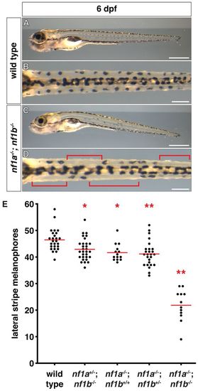
nf1 mutants display decreased lateral stripe melanophores. (A-D) Lateral stripe melanophores of wild-type (A,B) larvae demonstrate a normal uniform pattern that is absent in nf1a-/-; nf1b-/- larvae (C,D) at 6 dpf. The brackets in D highlight regions where melanophores are absent. (E) Quantification of lateral stripe melanophores from wild-type (n=26), nf1a+/-; nf1b-/- (n=30), nf1a-/-; nf1b+/+ (n=14), nf1a-/-; nf1b+/- (n=26) and nf1a-/-; nf1b-/- (n=12) larvae at 6 dpf. Each point represents the number of lateral stripe melanophores in an individual embryo and red lines indicate mean values. *P<0.05, **P<0.01. Scale bars: 0.5 mm (A,C) and 150 μm (B,D).
|