
FIGURE
Fig. 4
- ID
- ZDB-FIG-120627-20
- Publication
- Ton et al., 2012 - Semaphorin3d mediates Cx43-dependent phenotypes during fin regeneration
- Other Figures
- All Figure Page
- Back to All Figure Page
Fig. 4
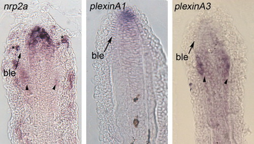
Gene expression of candidate receptors for Sema3d. Left: expression of nrp2a is primarily located in the distal blastema and in skeletal precursor cells. Staining of individual cells of the outer epithelial cells is also observed (N). Middle: expression of plxna1 is primarily in the distal blastema and in the distal cells of the basal epidermis. Right: expression of plxna3 is located in both the skeletal precursor cells and in the blastema. Arrows identify the basal layer of the epidermis (ble), arrowheads identify skeletal precursor cells. |
Expression Data
| Genes: | |
|---|---|
| Fish: | |
| Condition: | |
| Anatomical Terms: | |
| Stage: | Adult |
Expression Detail
Antibody Labeling
Phenotype Data
Phenotype Detail
Acknowledgments
This image is the copyrighted work of the attributed author or publisher, and
ZFIN has permission only to display this image to its users.
Additional permissions should be obtained from the applicable author or publisher of the image.
Reprinted from Developmental Biology, 366(2), Ton, Q.V., and Iovine, M.K., Semaphorin3d mediates Cx43-dependent phenotypes during fin regeneration, 195-203, Copyright (2012) with permission from Elsevier. Full text @ Dev. Biol.