
Fig. S6
- ID
- ZDB-FIG-120316-35
- Publication
- Liao et al., 2012 - Tol2 gene trap integrations in the zebrafish amyloid precursor protein genes appa and aplp2 reveal accumulation of secreted APP at the embryonic veins
- Other Figures
- All Figure Page
- Back to All Figure Page
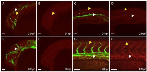
Confocal imaging of live appa+/is22Gt; Tg(flk1:moesin1-egfp)+/is1 embryo before completion of venous vessel formation. A–D: Lateral view, 24 hours post fertilization (hpf). A: Appa-red fluorescent protein (RFP) in the head is enriched in the eye surrounding the lens (yellow arrows). Appa-RFP has not accumulated at the veins in the head. C,D: Appa-RFP accumulates at the somite boundaries in the trunk (yellow arrow) but is absent from the posterior caudal vein region (white arrow). E–H: Lateral view, 28 hpf. E,F: Appa-RFP does not show enrichment at the veins in the head at this time. G,H: Accumulation of Appa-RFP at the somite boundaries has increased (yellow arrows) and is detected in the region of the posterior caudal vein (white arrows). Scale bars = 50 μm. |