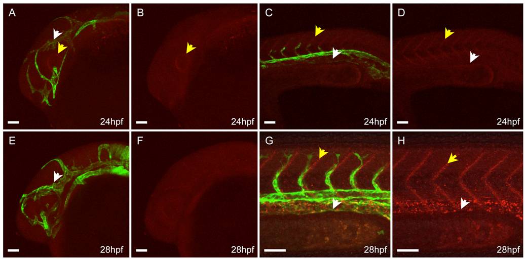
Fig. S6 Confocal imaging of live appa+/is22Gt; Tg(flk1:moesin1-egfp)+/is1 embryo before completion of venous vessel formation. A–D: Lateral view, 24 hours post fertilization (hpf). A: Appa-red fluorescent protein (RFP) in the head is enriched in the eye surrounding the lens (yellow arrows). Appa-RFP has not accumulated at the veins in the head. C,D: Appa-RFP accumulates at the somite boundaries in the trunk (yellow arrow) but is absent from the posterior caudal vein region (white arrow). E–H: Lateral view, 28 hpf. E,F: Appa-RFP does not show enrichment at the veins in the head at this time. G,H: Accumulation of Appa-RFP at the somite boundaries has increased (yellow arrows) and is detected in the region of the posterior caudal vein (white arrows). Scale bars = 50 μm.
                
                    
                        Image


                        Figure Caption


                        Acknowledgments


This image is the copyrighted work of the attributed author or publisher, and
ZFIN has permission only to display this image to its users.
Additional permissions should be obtained from the applicable author or publisher of the image.
      Full text @ Dev. Dyn.


        