
Fig. 2
- ID
- ZDB-FIG-120315-30
- Publication
- Clendenon et al., 2012 - Zebrafish cadherin-11 participates in retinal differentiation and retinotectal axon projection during visual system development
- Other Figures
- All Figure Page
- Back to All Figure Page
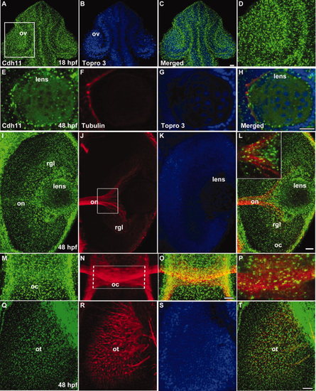
Distribution of Cdh11 in the developing lens, retina, and retinotectal projections. Laser scanning confocal microscope images are shown. Panels A–H, L (inset), and P are single optical sections, and other images are projection images of several optical sections to show neural projections through the tissues. Embryos at 18 hours postfertilization (hpf) were double labeled using antibodies that specifically recognize Cdh11 (green, panels A, C, and D) and TO-PRO-3 (nuclear stain, blue, panels B and C). Cdh11 and nuclear label images were merged (panel C). The boxed region in panel A contains the optic vesicle (ov), showing details of the retina and lens, are shown in panel D. A–D: Dorsal views are shown, and anterior is up. Embryos at 48 hours postfertilization (hpf) were triple labeled using antibodies that specifically recognize Cdh11 (green, panels E, I, M, and Q); antibodies that specifically recognize acetylated tubulin (red, panels F, J, N, and R); and TO-PRO-3 that labels nuclei (blue, panels G, K and S). Merged images (panels H, L, O, P, and T) are shown for comparison of expression patterns. Cdh11 was detected in the retina and lens at 18 and 48 hpf. E–H,M–T: Ventral views are shown, and anterior is up. I–L: Anterior is left, and lateral is up. Acetylated tubulin labels neuronal processes, including the optic nerve (on, panels J and L); the optic chiasm (oc, panels N, O and P); and the retinotectal projections arriving in the optic tectum (ot, panels R and T). Views of the eye (I and L) and brain (M, O, P, Q, and T) illustrate Cdh11 staining is associated with the optic nerve and optic chiasm. Scale bars = 20 μm. |
| Gene: | |
|---|---|
| Antibody: | |
| Fish: | |
| Anatomical Terms: | |
| Stage Range: | 14-19 somites to Long-pec |