
Fig. 3
- ID
- ZDB-FIG-120315-31
- Publication
- Clendenon et al., 2012 - Zebrafish cadherin-11 participates in retinal differentiation and retinotectal axon projection during visual system development
- Other Figures
- All Figure Page
- Back to All Figure Page
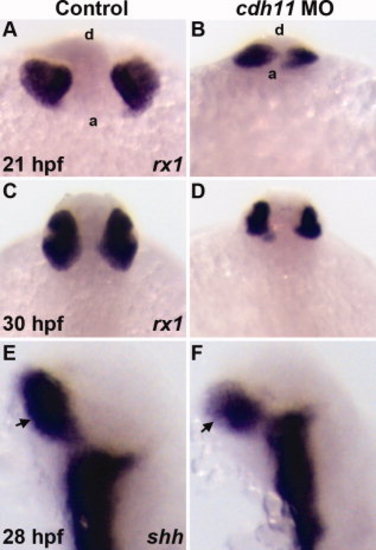
Expression of early retinal specification markers in cdh11 morphant embryos. Zebrafish embryos were injected with either standard control morpholino oligonucleotide (MO, Control) or cdh11 MO. Whole-mount in situ hybridization labeling of embryos at different stages was performed using probes for markers retinal specification. Probes are shown in the bottom right corner of each panel. A,B:rx1 labeling of 21 hours postfertilization (hpf) control and cdh11 morphant embryos. Anterior views from a ventral perspective; a, anterior embryo border; d, dorsal surface. rx1 labeling was detected in control (A) and cdh11 morphant embryos (B). C,D:rx1 labeling was strongly detected in 30 hpf control and cdh11 morphant embryos. Dorsal view of 30 hpf embryos: anterior is top. E,F: Expression of shh, a marker of retinal stalk (arrows) differentiation, was strongly detected in 28 hpf control and cdh11 morphant embryos; lateral views, anterior is up. |
| Fish: | |
|---|---|
| Knockdown Reagent: | |
| Observed In: | |
| Stage Range: | 20-25 somites to Prim-15 |