
Fig. 4
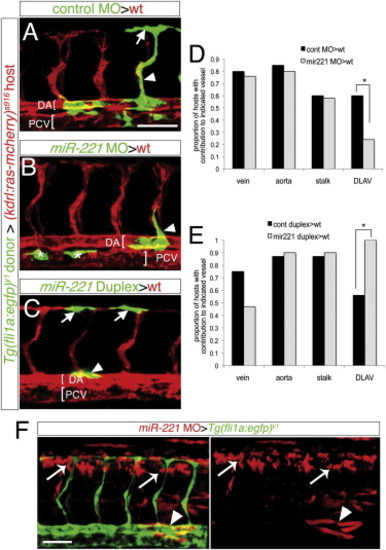
miR-221 Acts Endothelial Cell Autonomously to Drive Tip Cell Potential (A–C and F) Confocal micrographs of mosaic embryos at 27 hpf following cell transplantation. (A–C) Donor Tg(fli1a:egfp)y1 cells are green, host Tg(kdrl:ras-mcherry)s916 vessels are red; DA, dorsal aorta; PCV, posterior cardinal vein, both indicated by brackets. (A) Donor control cells in DLAV (arrow) and ISV stalk (arrowhead). (B) Donor miR-221-deficient cells in the ISV stalk (arrowhead). Asterisks indicate donor cells in the PCV. (C) Donor cells overexpressing miR-221 in DLAV (arrows) and dorsal aorta (arrowhead). (D and E) Proportion of host embryos with donor contribution to indicated blood vessel type. (D) -p = 0.04. (E) -p = 0.001. (F) Tg(fli1a:egfp)y1 host embryo with miR-221-deficient donor cells labeled with rhodamine in nonendothelial cell types surrounding the ISVs, including neural tube (white arrows) and somites (white arrowheads). Scale bars are 50 μm. |
| Gene: | |
|---|---|
| Fish: | |
| Anatomical Terms: | |
| Stage: | Prim-5 |
Reprinted from Developmental Cell, 22(2), Nicoli, S., Knyphausen, C.P., Zhu, L.J., Lakshmanan, A., and Lawson, N.D., miR-221 Is Required for Endothelial Tip Cell Behaviors during Vascular Development, 418-429, Copyright (2012) with permission from Elsevier. Full text @ Dev. Cell