
Fig. 7
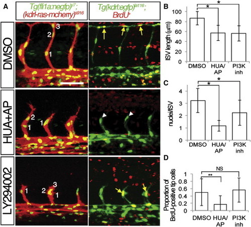
Proliferation and PI3K Are Required for ISV Growth (A) Embryos at 27 hpf treated with 4% DMSO, 150 μM 5-hydroxyurea, and 20 mM aphidocolin (HUA/AP), or 25 μM LY294002 (PI3K inh) beginning at 20 hpf. Left panels, confocal micrographs of Tg(fli1:negfp)y7;(kdrl:ras-cherry)s916 embryos. (right panels) Two-photon micrographs of Tg(kdrl:egfp)la116 embryos pulsed with BrdU at 20 hpf. Lateral views, dorsal is up, anterior to the left, yellow arrows denote BrdU-positive endothelial nuclei; BrdU-negative endothelial nuclei indicated by white arrowheads. Scale bar is 50 μm. (B and C) Quantification of (B) ISV length in microns and (C) number of nuclei per ISV in Tg(fli1:negfp)y7;(kdrl:ras-cherry)s916 embryos at 27 hpf treated as in (A). -p < 0.0001. (D) Quantification of BrdU-positive DLAV or ISV tip cells in Tg(kdrl:egfp)la116 embryos at 27 hpf treated as in (A). --p < 0.02. NS, not significant. |
| Fish: | |
|---|---|
| Conditions: | |
| Observed In: | |
| Stage: | Prim-5 |
Reprinted from Developmental Cell, 22(2), Nicoli, S., Knyphausen, C.P., Zhu, L.J., Lakshmanan, A., and Lawson, N.D., miR-221 Is Required for Endothelial Tip Cell Behaviors during Vascular Development, 418-429, Copyright (2012) with permission from Elsevier. Full text @ Dev. Cell