Fig. 4
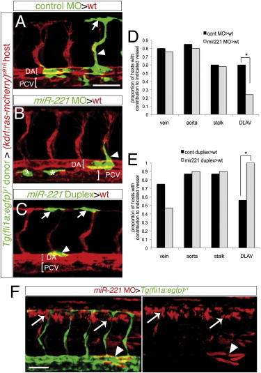
miR-221 Acts Endothelial Cell Autonomously to Drive Tip Cell Potential (A–C and F) Confocal micrographs of mosaic embryos at 27 hpf following cell transplantation. (A–C) Donor Tg(fli1a:egfp)y1 cells are green, host Tg(kdrl:ras-mcherry)s916 vessels are red; DA, dorsal aorta; PCV, posterior cardinal vein, both indicated by brackets. (A) Donor control cells in DLAV (arrow) and ISV stalk (arrowhead). (B) Donor miR-221-deficient cells in the ISV stalk (arrowhead). Asterisks indicate donor cells in the PCV. (C) Donor cells overexpressing miR-221 in DLAV (arrows) and dorsal aorta (arrowhead). (D and E) Proportion of host embryos with donor contribution to indicated blood vessel type. (D) -p = 0.04. (E) -p = 0.001. (F) Tg(fli1a:egfp)y1 host embryo with miR-221-deficient donor cells labeled with rhodamine in nonendothelial cell types surrounding the ISVs, including neural tube (white arrows) and somites (white arrowheads). Scale bars are 50 μm.
Reprinted from Developmental Cell, 22(2), Nicoli, S., Knyphausen, C.P., Zhu, L.J., Lakshmanan, A., and Lawson, N.D., miR-221 Is Required for Endothelial Tip Cell Behaviors during Vascular Development, 418-429, Copyright (2012) with permission from Elsevier. Full text @ Dev. Cell

