
Fig. 4
- ID
- ZDB-FIG-120216-85
- Publication
- Wu et al., 2011 - Ryanodine receptors, a family of intracellular calcium ion channels, are expressed throughout early vertebrate development
- Other Figures
- All Figure Page
- Back to All Figure Page
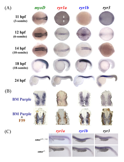
Zebrafish ryr1a, ryr1b and ryr3 mRNA is localised to embryonic skeletal muscle. (a) Expression of zebrafish myoD, ryr1a, ryr1b and ryr3 was examined using whole mount in situ hybridisation. Expression of ryr1a mRNA is detectable in the adaxial cells of 11 hpf embryos (3-somite, arrows), whereas ryr1b mRNA expression is present in cells adjacent to the notochord from 12 hpf (6-somite stage) embryos. Expression of ryr3 mRNA was only evident at 24 hpf, with the strongest staining observed in the anterior somites. The embryos at 11 and 12 hpf are orientated so that the anterior is to the left. (b) Cross-sections showing myoD, ryr1a, ryr1b and ryr3 in-situ mRNA hybridisation (above) and double immunostained with the F59 antibody/HRP labeling (below) in 24 hpf embryos. (c) ryr1a, ryr1b and ryr3 mRNA expression in wildtype (smo+/+), heterozygote (smo+/-) and homozygous (smo-/-) mutants at 24 hpf. There is no ryr1a mRNA expression in the smo-/- mutant, compared to wildtype and heterozygous embryos at 24 hpf. |
| Genes: | |
|---|---|
| Antibody: | |
| Fish: | |
| Anatomical Terms: | |
| Stage Range: | 1-4 somites to Prim-5 |