FIGURE
            Fig. S3
                
                    
                        Fig. S3


| 
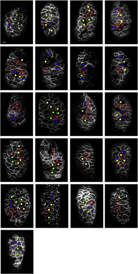
 Spatial distribution of behavioral heterogeneity in wild-type hearts. Images display the entire collection of wild-type hearts examined by live imaging. Lateral views, oriented with the arterial pole at the top, depict Tg(myl7:mkate-caax) expression in each ventricle at 40 hpf. Dots indicate all of the examined cardiomyocytes, and each dot′s color corresponds to the observed behavior of that cell, according to the color code established in Fig. 5. Scale bar is 10 μm. | 
                
                    
                        Expression Data


                        Expression Detail


                        Antibody Labeling


                        Phenotype Data


                        Phenotype Detail


                        Acknowledgments


This image is the copyrighted work of the attributed author or publisher, and
ZFIN has permission only to display this image to its users.
Additional permissions should be obtained from the applicable author or publisher of the image.
    Reprinted from Developmental Biology, 362(2), Lin, Y.F., Swinburne, I., and Yelon, D., Multiple influences of blood flow on cardiomyocyte hypertrophy in the embryonic zebrafish heart, 242-253, Copyright (2012) with permission from Elsevier. Full text @ Dev. Biol.
 
        