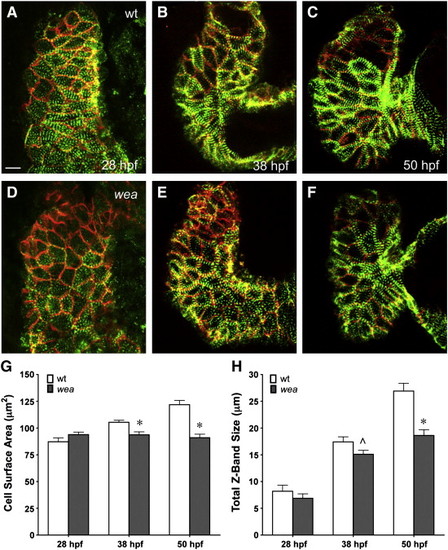
Fig. 6
|
Myofibril maturation is diminished in wea mutant embryos. (A–F) Ventral views, arterial pole at the top, of dissected hearts at 28, 38, and 50 hpf depict localization of Actn3b-egfp (green) in Z-bodies and Z-bands and Dm-grasp (red) at cell boundaries. Actn3b-egfp and Dm-grasp localization indicate the dynamic progression of cell size and myofibril content during chamber emergence. Wild-type (wt) and wea mutant ventricular cardiomyocytes are indistinguishable at 28 hpf (A, D), but cell size expansion and myofibril growth and organization do not progress normally in wea mutants (B, C, E, F). Scale bar is 10 μm. (G) Bar graph indicates mean cell surface area measurements in μm2 for ventricular cardiomyocytes at the indicated stages; error bars indicate standard error. Asterisks indicate statistically significant differences between wild-type and wea (p < 0.05); these differences become apparent by 38 hpf. (H) Bar graph indicates mean measurements of total Z-band size in μm per ventricular cardiomyocyte at the indicated stages; error bars indicate standard error. The asterisk indicates a statistically significant difference between wild-type and wea at 50 hpf (p < 0.05). The carat indicates a less striking difference between wild-type and wea at 38 hpf (p = 0.06). |
Reprinted from Developmental Biology, 362(2), Lin, Y.F., Swinburne, I., and Yelon, D., Multiple influences of blood flow on cardiomyocyte hypertrophy in the embryonic zebrafish heart, 242-253, Copyright (2012) with permission from Elsevier. Full text @ Dev. Biol.

