
Fig. S2
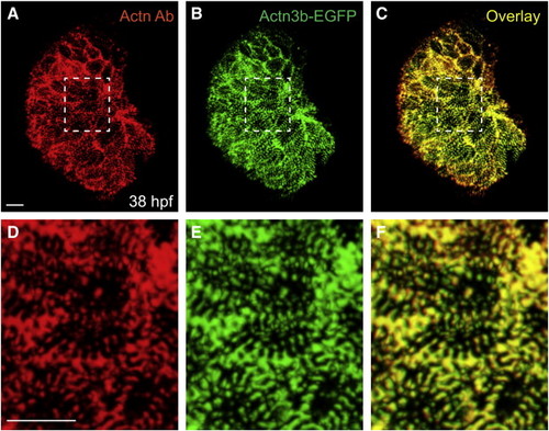
Tg(myl7:actn3b-egfp) faithfully labels myofibril Z-bands and Z-bodies. (A–C) Ventral view, arterial pole at the top, of a ventricle at 38 hpf from an embryo expressing Tg(myl7:actn3b-egfp). (A) Immunofluorescence (red) localizes α-actinin in Z-bands and Z-bodies within ventricular cardiomyocytes. (B) Native green fluorescence indicates incorporation of the Actn3b-egfp fusion protein into Z-bands and Z-bodies. (C) Overlay and comparison of red and green fluorescence indicates strong correspondence between the incorporation of Actn3b-egfp and the localization of α-actinin. Therefore, the Tg(myl7:actn3b-egfp) transgene provides an effective method for labeling myofibril Z-bands and Z-bodies. (D–F) Higher magnification views of the regions marked by white dashed rectangles in panels A–C. Scale bars are 10 μm. |
Reprinted from Developmental Biology, 362(2), Lin, Y.F., Swinburne, I., and Yelon, D., Multiple influences of blood flow on cardiomyocyte hypertrophy in the embryonic zebrafish heart, 242-253, Copyright (2012) with permission from Elsevier. Full text @ Dev. Biol.