
Fig. 4
- ID
- ZDB-FIG-120130-4
- Publication
- Schuhmacher et al., 2011 - Evolutionary relationships and diversification of barhl genes within retinal cell lineages
- Other Figures
- All Figure Page
- Back to All Figure Page
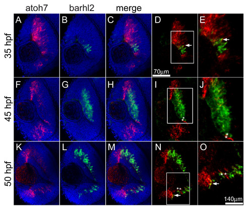
Double fluorescent in situ hybridization of barhl2 and atoh7. Confocal sections through the central retina of embryos hybridized with barhl2 (revealed with FITC, shown in green) and atoh7 (revealed with Cy3, shown in red), antisense RNA probes. Nuclei were stained with DAPI (blue) to outline retinal layers. View is frontal in all pictures, anterior is always to the top. (D-E and N-O) White arrows show co-localization of both mRNAs in cells at the border of the expression domains. (I-J and N-O) white asterisks indicate adjacent cells expressing either barhl2 or atoh7. (D, I and N) white squares highlight the magnified area in E, J and O, respectively. |
| Genes: | |
|---|---|
| Fish: | |
| Anatomical Terms: | |
| Stage Range: | Prim-15 to Long-pec |