
Fig. 1
- ID
- ZDB-FIG-120130-1
- Publication
- Schuhmacher et al., 2011 - Evolutionary relationships and diversification of barhl genes within retinal cell lineages
- Other Figures
- All Figure Page
- Back to All Figure Page
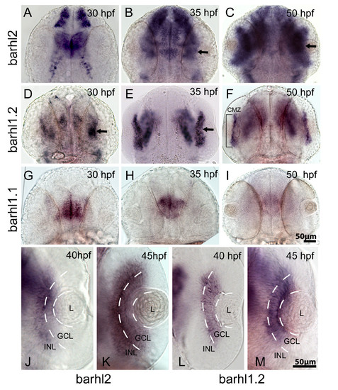
Comparative in situ hybridization of barhl paralogs expression in the zebrafish retina. Dorsal view of wild-type zebrafish embryos hybridized with barhl2 (A-C, J-K), barhl1.2 (D-F, L, M) and barhl1.1 (G-I), antisense RNA probes. Stages analyzed are indicated. Anterior is always to the top. (B, C, D and E) black arrows in indicate expression localized in the retina. In (F), the black bracket highlights barhl1.2 expression restricted in a thin retinal domain, which is the ciliary marginal zone (CMZ). (J-M) show a closer view on individual retinas. White dashed lines highlight the boundary between lens (L), ganglion cell layer (GCL) and inner part of the inner nuclear layer (INL), where ACs are located. |
| Genes: | |
|---|---|
| Fish: | |
| Anatomical Terms: | |
| Stage Range: | Prim-15 to Long-pec |