
Fig. 8
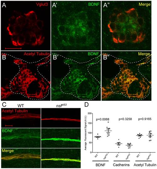
Expression of BDNF in wild-type hair cells and the pLLG, and accumulation of BDNF protein in the lateral line nerve of nsfst53 mutants. A, A single optical section of a neuromast from a wild-type zebrafish larva (5 dpf) immunolabeled for Vglut3 (A, red), BDNF (A′, green), and merged in (A′′). B, A single optical section of a wild-type pLLG, immunolabeled for acetylated Tubulin (B, red), BDNF (B′, green), and merged in (B′′). The cell bodies of the pLLG are outlined by dash lines. C, Representative z-projections of 6 sections (1 μm each) of acetylated Tubulin (red) and BDNF (green) immunolabeled lateral line nerves in wild-type and nsfst53 mutant larvae. D, The average pixel intensity (A.U.) of BDNF (WT, n = 7; nsfst53, n = 8), PAN-Cadherin (WT, n = 8; nsfst53, n = 5), and acetylated Tubulin labeling (WT, n = 7; nsfst53, n = 8) in wild-type control and nsfst53 mutants. Scale bar is 10 μm across all panels. |
| Antibodies: | |
|---|---|
| Fish: | |
| Anatomical Terms: | |
| Stage: | Day 5 |