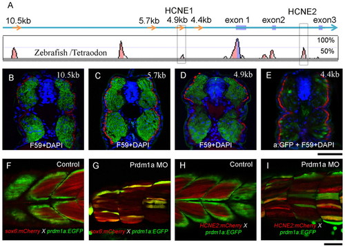
Conserved cis-elements recapitulate the sox6 expression pattern in the myotome. (A) Conserved non-coding elements (represented by peaks) upstream and downstream of the transcription start site revealed by alignment of the zebrafish and Tetraodon sox6 genomic sequences. Several of these are also highly conserved in mammals (see Fig. S3 in the supplementary material), including HCNE1 and HCNE2 (red boxes). (B-E) Transverse sections through mid-trunk regions of 48 hpf embryos transgenic for sox6 reporter genes; slow-twitch fibres are visualised with mAb F59 (red), nuclei by DAPI staining (blue). The 10.5 kb, 5.7 kb and 4.9 kb fragments drive bright and specific EGFP (green) expression exclusively in fast-twitch fibres; by contrast, the 4.4 kb fragment drives low-level mosaic expression of EGFP, detectable only in fixed specimens using anti-EGFP antibody (E). (F-I) Optical sagittal sections of 48 hpf Tg(PACprdm1:GFP)i106 embryos carrying the sox6 5.7 kb or HCNE2 mCherry reporter constructs. Both constructs are ectopically expressed (red) in fibres derived from adaxial cells (green) following injection of Prdm1a morpholino (G,I). Scale bars: 50 μm.
|
